Hyaluronic acid (HA), also known as hyaluronan, is a biocompatible, naturally occurring, unbranched glycosaminoglycan consisting of N-acetyl-D-glucosamine and D-glucuronic acid. This polymer is present in various tissues, including embryonic, epithelial, connective, and neural tissues. Due to its negative charge, HA is highly hydrophilic and plays a role in tissue structure and hydration. HA acts as an essential component of the extracellular matrix (ECM) and performs a variety of biological functions such as cell proliferation, migration, and signal transduction. The main producers are mesenchymal cells, astrocytes in the central nervous system, and fibroblasts in the heart. In human skin, about one-third of HA is renewed daily.
Mechanisms of hyaluronic acid action
HA acts both passively as a structural molecule through tissue hydration and stabilization of the extracellular space, and actively as a signaling molecule that modulates pro- and anti-inflammatory activities, cell migration, cell division, and differentiation.
Hyaluronic acid receptors and their functions
HA interacts with various HA-binding proteins and receptors such as CD44, RHAMM, LYVE-1, HARE, and Layilin. These receptors are involved in signaling pathways that control cell activation and movement.. CD44 is a multifunctional transmembrane glycoprotein expressed in almost all human cell types. It is involved in cell adhesion, migration, ECM turnover, and signal transduction. CD44 interacts with the cytoskeleton and is overexpressed in many solid tumors, where it promotes cell migration, proliferation, and invasion. RHAMM (CD168) exists in different isoforms and localizations (membrane, cytoplasm, nucleus). Extracellular RHAMM promotes cell migration.
Role of hyaluronic acid in development, wound healing, and cancer
HA is central to embryonic development, morphogenesis, tissue renewal, and wound healing. It supports cell migration, regulates inflammatory responses, and promotes tissue reconstruction. In tumors, HA is often upregulated, supporting cell proliferation, preventing apoptosis, facilitating nutrient diffusion, and promoting cell migration. CD44 and RHAMM play an important role in regulating cell motility and signaling pathways that promote tumor progression.
Breakdown of hyaluronic acid
Breakdown of HA occurs mainly through hyaluronidases (Hyal1, Hyal2, Hyal3, PH-20) and oxygen species (ROS) generated by many types of cells under stressed conditions. Hyal1 and Hyal2 are the most common and ubiquitous hyaluronidases and present in almost all somatic tissues.
Hyaluronic acid is introduced into 3-D Life Hydrogels through the crosslinkers HyLink and CD-HyLink. The hyaluronic acid in HyLink and CD-HyLink is of non-animal origin and obtained from microbial fermentation.
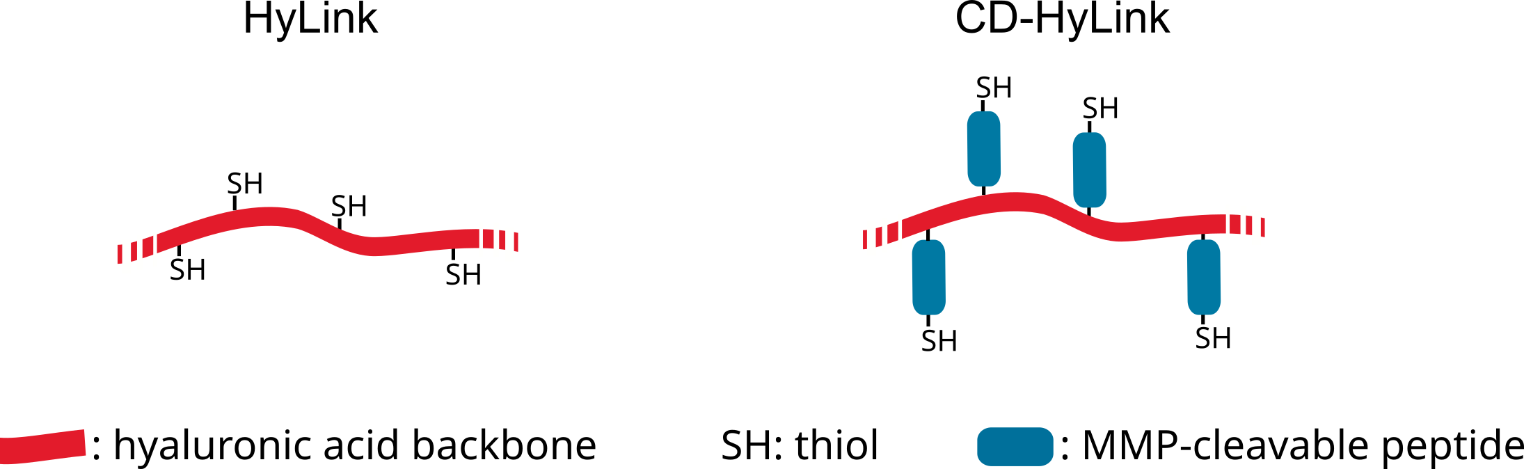
HyLink is composed of hyaluronic acid modified with thiol groups along the backbone of the molecule. In comparison, CD-HyLink additionally carries matrix metalloproteinase (MMP)-cleavable peptides between the molecule backbone and the thiol groups (Fig.1).
Figure 1: Schematic drawing of structure of HyLink and CD-HyLink.
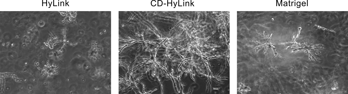
Both crosslinkers can be digested by hyaluronidases. In addition to the digestion by hyaluronidases, CD-HyLink can also be digested by MMPs due to the MMP cleavage sites. The combination of hyaluronidase and MMP sensitivity of CD-HyLink allows an enhanced cell spreading and migration when compared with HyLink at comparative hydrogel stiffness and also surpasses cell spreading and migration observed in Matrigel (Fig.2).
Figure 2: 3T3 cells were cultured for 5 days in biomimetic hydrogels consisting of SG-Dextran crosslinked either with HyLink or CD-HyLink (Dextran-HA Hydrogel and Dextran CD-HA Hydrogel, respectively) at a crosslinking strength of 1.2 mmol/L reactive groups. In addition, hydrogels had been both modified with 0.5 mmol/L of a cell adhesion peptide containing the RGD motif (RGD Peptide).